INTERNAL IMPINGEMENT

Constellation of symptoms which result from the greater tuberosity of the humerus and the articular surface of the rotator cuff abutting the posterosuperior glenoid when the shoulder is in an abducted and externally rotated position.

    • (Walch G, Boileau P, Noel E, Donell ST. Impingement of the deep surface of the supraspinatus tendon on the posterosuperior glenoid rim: An arthroscopic study. J Shoulder Elbow Surg1992)

History

  • Bennett, 1950 – secondary to inflammation in the posterior capsule and inferior glenohumeral ligament due to triceps traction.
  • Exostosis of the posteroinferior glenoid rim, which became known as the “Bennett lesion”.
  • Also described the presence of articular-sided posterosuperior rotator cuff tears.
  • In 1977, Lombardo et al further described ossification of the posterior glenoid rim
  • In 1985, Andrews et al described a series of overhead athletes with posterior shoulder pain that developed articular-sided posterosuperiorcuff tears with concomitant SLAP lesions
  • Jobeet al reported on a series of overhead athletes with posterosuperior impingement associated with anterior instability
  • In 1992, Walchet alreported a series of patients with impingement between the articular side of the supraspinatus tendon and the posterosuperior edge of the glenoid cavity, typically noted during maximal abduction and external rotation
  • The undersurface of the posterior rotator cuff becomes entrapped between the labrum and the greater tuberosity in the abductionexternal rotation position.

Pathophysiology and biomechanics

  • Multifactorial : physiologic shoulder remodelling posterior capsular contracture scapulardyskinesis.
  • The kinetic chain concept describes the coordinated motion that transmits energy in a synchronized fashion from the lower extremity, through the trunk, to the shoulder
  • Inflexibility, weakness, and imbalance of any point in the kinetic chain can create a situation where the arm lags behind the legs and trunk, placing the throwing shoulder, increasing stresses about the shoulder and leading to injury
  • Arthrosopy , 2003
  • JSES,2000

Throwing

6 phases

  • wind-up
  • early cocking
  • late cocking phase – abduction and max external rotation (internal impingement)
  • acceleration phase – greatest angular velocity (7200*/sec)
  • deceleration phase – shearing forces on labrum
  • follow through phase

Thrower’s paradox

  • The arc of motion in a high-level throwing athlete is shifted posteriorly to allow for increased external rotation at the cost of decreased internal rotation by allowing increased clearance of the greater tuberosity over the glenoid during rotation
  • increased retroversion of the humeral head and glenoid
  • increased anterior capsular laxity adaptations
  • Jobeet al originally described “subtle instability”, or microinstability, to define the acquired laxity and anterior translation of the humeral head.
  • Despite the need for increased laxity, adequate stability must be maintained to prevent symptomatic humeral head subluxation, often achieved through further posterior capsular contracture.
  • Grossman et al, who reported on a simulated posterior capsular contraction model which led to GIRD and posterosuperior translation of the humeral head during the late cocking, ultimately resulting in SLAP injuries.
  • Burkhart et al, have reported that shoulders with an internal rotation deficit > 25 degrees are at increased risk for development of SLAP lesions as a result of increased posterosuperior peel back on the labrum.

Scapular dyskinesis

  • When the scapula is ineffective in stabilizing the shoulder, the rotator cuff is forced to over-compensate to stabilize the glenohumeral joint.
  • These loads are then transmitted to the superior glenoid and the articular surface of the rotator cuff tendons and can lead to injury.
  • An independent factor in the development of internal impingement.
  • scapular dyskinesis has been reported in up to 100% of patients with internal impingement

Clinical features

  • shoulder stiffness
  • the need for a prolonged warm
  • note a decline in performance, including loss of control or decreases in pitch velocity
  • posterior shoulder pain, especially in the late cocking phase.


  • Jobe defined three stages in the clinical presentation of internal impingement.
  • Stage Ⅰ: consists of stiffness and difficulty in warming up, but no complaints of pain.
  • StageⅡ: hallmarked by the complaint of pain during the late cocking phase of the throwing cycle.
  • Stage Ⅲ : Those patients that have recurrent pain after a period of adequate rest and rehabilitation

Physical examination

  • Posterior joint line tenderness +
  • Incresed ER and decreased IR (in 90deg abd.)
  • The scapulae are evaluated for positioning, dyskinesis, and winging.
  • Rotator cuff : weakness in infraspinatus ( decreased ER strength)

SPECIAL TESTS

  • Relocation test : in 90deg abd– max ER Humerus pushed – anteriorly and inferiorly decrease pain
  • Posterior impingement sign : in 90 degabd, extension and max ER – deeper pain
  • Thrower’s paradox : they may exhibit anterior capsular laxity but they don’t have any apprehension ( in pathological damage +)
  • Posterior labrum and superior labrum : o’breintest 

Radiology

  • X ray :
  • “Bennett lesion” (exostosis of the posteroinferior glenoid rim)
  • sclerosis of the greater tuberosity
  • posterior humeral head osteochondral cysts
  • rounding of the posterior glenoid rim

AJR,2005

  • Mri :
  • Posterosuperior labral tears
  • partial-thickness articular-sided rotator cuff tears most notably at the junction between the supraspinatus and infraspinatus as they insert on to the humeral head
  • cystic changes in the posterior aspect of the humeral head
  • calcification at the scapular attachment of the posterior capsule (Bennett lesion)
  • posterior capsular contracture and thickening at the level of the posterior band of the inferior glenohumeral ligament
  • subchondral fracture and remodeling of the posterosuperior glenoid.


ARTHROSCOPY


Treatment

  • Conservative  :
  1. Rest for 4-6 weeks
  2. NSAIDS
  3. Physiotherapy : both treatment and preventive SLEEPER’S STRETCH : improves IR
  4. Scapular dyskinesis : by improving / strengthening of KINETIC chain (from lower body to sholder)

  • Operative :
  1. Arthroscopic posterior capsular release
  2. debridement or repair of partial tears
  3. Labral repair
  4. Osteotomy of humerus– increase retroversion
  5. Sonnery-Cottetet al performed arthroscopic debridement on twenty-eight tennis players with articular-sided partial tears and glenoid lesions.
  6. Seventy-nine percent were able to return to play, but 91% still had some persistent pain.
  7. If partial tear completion and repair is indicated, a lateralized double-row repair as described by Dines et al, should be considered as it has shown favorable outcomes in professional overhead throwing athletes, allowing restoration of a more anatomic footprint.
  8. Riandet al reported on humeral osteotomies to increase humeral retroversion in twenty patients who had continued pain after arthroscopic debridement.

  • SPORTS MEDICINE, 2014

Q&As on Shoulder Internal Impingement

1) What Is Shoulder Internal Impingement?

Shoulder discomfort in overhead athletes (football or volleyball players) is often brought on by internal impingement. Shoulder impingement happens when your rotator cuff rubs up against (or impinges on) or is pinched below  by the upper outer edge of your shoulder blade, known as the acromion. When the arm is put in extreme ranges of abduction and external rotation, the larger tuberosity of the humeral head’s posterior side repeatedly or excessively contacts the posterior-superior aspect of the glenoid border, causing the condition. In the end, this causes the supraspinatus and infraspinatus rotator cuff tendons and the glenoid labrum to impinge. Anterosuperior and posterosuperior are the two forms of internal impingement. . However, anterosuperior impingement rarely occurs.

2) What Causes the Development of Shoulder Impingement Conditions?

Similar to how your ankle expands after getting sprained, your rotator cuff does the same when inflamed or damaged. However, since your rotator cuff is encircled by bone, swelling results in different outcomes. Swelling of the rotator cuff narrows the area surrounding it, causing it to press on the acromion. The rotator cuff tendons’ friction causes swelling, further reducing the available space below the acromion, creating a vicious cycle. As the region where the rotator cuff resides becomes even more constrained due to bone spurs on the acromion bone, impingement may occur.

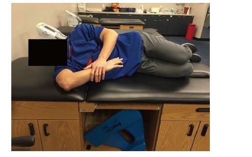
3) What Are the Symptoms of Internal Impingement?

Shoulder impingement condition symptoms involve:

  • Discomfort when your arms are raised over your head.
  • Pain while raising, lowering  after being elevated, or extending your arm.
  • Your front shoulder may be sore and painful.
  • Shifting shoulder pain that radiates down the side of your arm.
  • Pain while lying on the troubled side.
  • Your ability to sleep is hampered by nighttime pain or aches in the shoulder.
  • Experiencing discomfort while reaching behind your back 
  • Weakness and stiffness in the shoulders and arms.

4) What Are the Non-surgical Treatments for Internal Impingement?

Early on and when symptoms are modest, conservative therapy consisting of rest and activity restriction,  non-steroidal anti-inflammatory drugs (NSAIDs) may also be beneficial.

Physiotherapy: To extend the joint and avoid mobility loss, sessions of a rehabilitation programme with  a physical or occupational therapist may help.. Expectations and the degree of suffering influence success.

Injections: An injection in the AC joint that is guided by ultrasonography or fluoroscopy results in a reduction of pain and inflammation.

While the benefits of cortisone are often transient, they may provide quite effective pain relief in the short term. It also helps to support the diagnosis.

5) What Is an Arthroscopic Technique for Internal Impingement?

If non-operative therapy is unsuccessful, one has to opt for a surgical procedure; generally, laparoscopic surgery is the way to go.. In laparoscopy, instead of making a major incision, a small incision is used to insert a fiber-optic scope and a tiny, pencil-sized equipment. The surgeon shall repair the issue by looking at the area via a video feed thanks to the arthroscope’s connection to a television display.

After surgery, the arm will have to spend a brief time in a sling. This’ll enables quick healing. The sling may be removed as soon as one feels comfortable to start exercising and using the arm.

© Copyright 2022 Bangalore Shoulder Institute